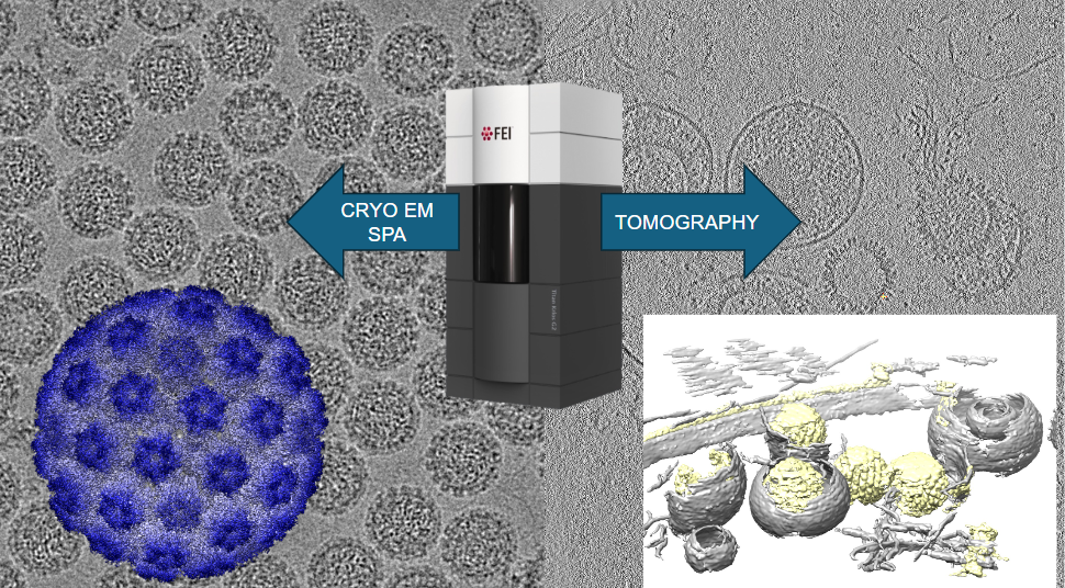
Cryo EM is used to create high resolution 3-dimensional models of biological materials which are too small to be seen in an optical microscope.

For single particle analysis, purified proteins or protein complexes are flash frozen in a thin layer of vitreous ice on a small grid which is inserted into the Cryo TEM microscope.

Large numbers of images of the particles in many different orientations within the ice are collected. Computational processing of these images allows the creation of 3-dimensional models which can show the location of atoms in the molecules being studied.

The details of these high resolution models can be used to study things such as how biological molecules function, how they bind to other molecules, how mutations can cause changes in function, and how drugs can interact with the biological molecules. These projects can lead to better understanding of the causes of diseases and help in the search for potential cures.

Cryo EM facilities, equipment, and computational resources are expensive to obtain and maintain, but having these resources available to University of Minnesota researchers can enhance their work. There are a few national cryo EM centers in the United States, but access to them involves submitting a proposal with preliminary results, waiting in a long queue for data collection, and traveling or shipping samples for long distances.
The Hormel Institute Cryo EM facility is more conveniently located and will enable researchers to screen and optimize their cryo-EM sample preparation, and collect high quality data without leaving Minnesota.