Cancer Biology
Cancer Biology
Luke H. Hoeppner, PhD
Associate Professor
Our “Cancer Biology” research section studies the molecular mechanisms and signal transduction pathways involved in vascular permeability, cancer progression/metastasis, cancer drug resistance, and adverse effects of cancer therapy. We employ two talented postdoctoral fellows, Sk. Kayum Alam, Ph.D. and Li Wang, M.D., Ph.D. Over each of the past three summers, Christina Hernandez (2018), Abbygail Coyle (2017), and Erin Dankert (2016) have joined us as summer undergraduate research experience (SURE) interns. They eagerly learned a variety of technical laboratory skills and contributed to aspects of numerous research projects. Our National Institutes of Health funded laboratory was established in the fall of 2015.
Lung cancer is the leading cancer related cause of death in the United States and worldwide. Non-small cell lung cancer (NSCLC) represents 85% of all lung cancer and carries a very poor survival rate: less than 15% of patients survive more than five years. Despite administration of front-line chemotherapeutic agents with molecular targeted systemic therapies, the survival rate of NSCLC patients remains dismal due to the large number of individuals diagnosed with advanced stage disease and the primary and secondary resistance to current therapies. Elucidation of new biomarkers and novel precision therapies will help overcome these challenges and make significant strides in improving lung. The primary focus of our research is to develop new therapies to inhibit lung cancer progression and prevent tumor cells from acquiring resistance to current treatments. We accomplish these goals through the use of a variety of models as well as utilization of lung cancer patient samples. Such translational studies will be important for the development of new cancer therapies. Our research aims to improve the dismal survival rate of lung cancer patients and seeks an innovative approach to combatting tumor drug resistance. We seek to identify novel therapeutic targets to help the multitude of Americans suffering from lung cancer.
Vascular endothelial growth factor (VEGF) is required for blood vessel formation and promotes permeability in veins. Tumors produce VEGF because they require their own vasculature to grow, obtain nutrients and oxygen, and eliminate waste products. The permeability induced by VEGF enables cancer cells to escape their primary site, enter the bloodstream, and metastasize to other tissues. Dopamine (DA) and dopamine D2 receptor (D2R) agonists inhibit VEGF-mediated blood vessel development (angiogenesis) and vascular permeability by inhibiting VEGF binding, VEGF receptor phosphorylation and subsequent downstream signaling. Our recent studies demonstrated D2R agonists, including FDA approved cabergoline, inhibit lung cancer growth in in vivo models by reducing angiogenesis and tumor infiltrating immune suppressor cells. Pathological examination of human lung cancer tissue revealed a positive correlation between endothelial D2R expression levels and tumor stage as well as patient smoking history.
- Triggering the dopamine pathway to inhibit lung cancer progression
Currently, we are focused on understanding how a signaling molecule downstream of the dopamine D2 receptor, called DARPP-32 (dopamine and cyclic-AMP-regulated phosphoprotein), can be modulated to inhibit lung cancer growth. Recent studies have demonstrated that elevated expression of DARPP-32 and its truncated splice-variant, t-DARPP, are associated with breast and gastric tumorigenesis. Thus, we hypothesized DARPP-32 and t-DARPP proteins may activate oncogenic signaling that contributes to progression of NSCLC. We demonstrate that overexpression of DARPP-32 and t-DARPP promotes lung tumor growth in human xenograft orthotopic murine models through activation of AKT and ERK signaling. Correspondingly, abrogation of DARPP-32 in human NSCLC cells reduces lung tumor growth in preclinical in vivo models. We identify migration as a cellular mechanism by which DARPP-32 proteins stimulate NSCLC. Our studies suggest a novel physical interaction between DARPP-32 and inhibitory kappa B kinase-α (IKKα) promotes NSCLC cell migration through nuclear factor kappa-light-chain-enhancer of activated B cells 2 (NF-KB2) signaling via the non-canonical p52 pathway. Histopathological analysis of over 60 lung adenocarcinoma patientderived tissues reveals t-DARPP protein overexpression positively correlates with tumor grade, suggesting upregulation of t-DARPP promotes lung adenocarcinoma progression. Evaluation of bioinformatics data corresponding to over 500 lung adenocarcinoma patients confirms elevated t-DARPP expression is associated with worsening tumor grade as well as demonstrates correlation between high t-DARPP levels and poor overall NSCLC patient survival. Taken together, we describe aberrant DARPP-32 isoform expression promotes NSCLC growth and association of DARPP-32 with IKKα stimulates lung cancer cell migration via non-canonical NF-KB2 signaling. Identification of DARPP-32 signaling as a new potential molecular target for NSCLC therapy offers promise to improve the clinical outcome of patients afflicted with lung cancer.
- Development of zebrafish models of vascular permeability and cancer metastasis
VEGF induces vascular permeability in stroke, heart attack, and cancer leading to many pathophysiological consequences. Following cerebral or myocardial infarction, VEGF induces gaps between adjacent endothelial cells in ischemic tissue and the resulting vessel leakiness causes deleterious edema formation and tissue damage. In cancer, VEGF-mediated permeability promotes tumor angiogenesis and metastasis. The molecular mechanisms by which VEGF acts to induce hyperpermeability are poorly understood and in vivo models that easily facilitate real-time, genetic studies of permeability do not exist. We developed a heat-inducible VEGF transgenic zebrafish model through which vascular permeability can be monitored in real-time. Using this approach with protein knockdown, as well as knockout in vivo models, we described a novel role of phospholipase Cβ3 (PLCβ3) as a negative regulator of VEGF-mediated vascular permeability by tightly regulating intracellular calcium release. We have also used this zebrafish model to elucidate the role of RhoC and other molecules in vascular homeostasis. The zebrafish vascular permeability model represents a straightforward method for identifying genetic regulators of VEGF-mediated vascular as promising targets for cancer, heart disease and stroke therapies. We also developed a zebrafish xenograft model of human cancer cell metastasis, which has been used in two separate studies to support our findings from murine cancer models. We are currently using these models to elucidate the molecular regulation of vascular permeability and cancer metastasis.
- Topical treatment of radiotherapy-induced skin damage in breast cancer patients
Over three million women living in the United States have a history of invasive breast cancer. In 2017, an estimated 252,710 new cases of invasive breast cancer will be diagnosed and over 40,000 American women will unfortunately die due to this dismal disease. About half of all breast cancer patients in the United States receive radiation therapy. Radiotherapy uses targeted, high energy X-rays to effectively destroy cancer cells, but healthy skin tissue is also damaged in the process. Radiation-induced skin damage typically manifests as radiation dermatitis, a prevalent side effect affecting approximately 95% of radiotherapy recipients. The effects of radiation dermatitis include pain, itching, poor aesthetic appearance, and chronic reappearance of skin wounds due to pathological changes during the healing process, such as excessive fibrosis. Severe radiation dermatitis occurs in 5-10% of breast cancer patients receiving whole-breast radiotherapy, which leads to delays or stoppage of radiotherapy and increases the risk for cancer recurrence. Common approaches to prevent and reduce radiation-induced dermatitis of the irradiated skin area involve basic moisturizing, cleansing with mild soap, applying topical cortisone creams, and avoiding irritants like scratching or rough clothing. However, reports from a multitude of clinical trials conclude all current radiation dermatitis treatment strategies lack clinical efficacy. Therefore, new treatments that promote recovery from radiation dermatitis are necessary to improve the quality of life and clinical outcome of breast cancer patients by alleviating painful short- and long-term radiation side effects to ensure completion of radiation therapy regimens. The goal of our “Paint the Town Pink” supported work is to define the molecular mechanisms through which radiation causes skin injury and develop a topical treatment for radiation dermatitis. To achieve this aim, we are conducting ongoing studies using an in vivo model of radiotherapy-induced skin injury we developed in collaboration with UMN colleagues.
Cancer Biomarkers and Drug Resistance
Cancer Biomarkers and Drug Resistance
Ann Bode, PhD
Professor
Cancer is one of the leading causes of human death worldwide. By focusing on molecular mechanisms, we continue to discover the key molecular events in cancer development as well as agents for cancer prevention and therapy.
1. Discovery of key molecular events in cancer development.
a. The discovery of circulating prostaglandin biosynthesis in colorectal cancer. We found that the prostaglandin thromboxane A2 (TXA2) level is unexpectedly correlated with colorectal cancer progression. The TXA2 pathway is constitutively activated during colorectal tumorigenesis and required for anchorage-independent growth of colon cancer cells. Our work lays the foundation for introducing a TXA2-targeting strategy for the prevention, early detection and therapy of colon cancer. We further discovered that human colorectal cancer progression is accompanied by an elevation in epidermal growth factor receptor (EGFR) levels. These high levels of EGFR can be attenuated by aspirin intake. The widespread over-expression of EGFR occurs as a consequence of COX-2 activation in familial adenomatous polyposis (FAP) patients. This study revealed a functional association between COX-2 and EGFR expression during colon carcinogenesis and provided new strategies for colon cancer prevention and therapy. b. Discovery of novel mechanisms of RNF2 in cell death. RNF2, also known as Ring1B/Ring2, is a component of the polycomb repression complex 1. RNF2 is highly expressed in many tumors, suggesting it might have an oncogenic function, but the mechanism of action is unknown. We showed that knocking down RNF2 expression significantly inhibits both cell proliferation and colony formation in soft agar and induces apoptosis in cancer cells. Knockdown of RNF2 in HCT116 p53+/+ cells resulted in significantly more apoptosis than was observed in RNF2 knockdown HCT116 p53./. cells, indicating that RNF2 knockdown-induced apoptosis is at least partially, dependent on p53. Various p53-targeted genes were increased in RNF2 knockdown cells. Further studies revealed that in RNF2 knockdown cells, the p53 protein level was increased, the half-life of p53 was prolonged, and p53 ubiquitination was decreased. In contrast, cells over-expressing RNF2 showed a decreased p53 protein level, a shorter p53 half-life, and increased p53 ubiquitination. Importantly, we found that RNF2 directly binds with both p53 and MDM2 as well as promotes MDM2-mediated p53 ubiquitination. RNF2 over-expression also could increase the half-life of MDM2 and inhibit its ubiquitination. The regulation of p53 and MDM2 stability by RNF2 also was observed during the etoposide-induced DNA damage response. These results provide a possible mechanism explaining the oncogenic function of RNF2, and, because RNF2 is important for cancer cell survival and proliferation, it might be an ideal target for cancer therapy or prevention.
2. Discovery of novel targets and agents for skin cancer prevention and therapy.
Solar UV (SUV) irradiation is a major factor in skin carcinogenesis, the most common form of cancer in the United States. The mitogen-activated protein kinase (MAPK) cascades are activated by SUV irradiation. We found that p38 signaling is critical for skin carcinogenesis. The 90 kDa ribosomal S6 kinase (RSK) and mitogen and stress-activated protein kinase (MSK) proteins constitute a family of protein kinases that mediate signal transduction downstream of the MAPK cascades. Phosphorylation of RSK and MSK1 was upregulated in human squamous cell carcinoma (SCC) and SUV-treated mouse skin. Kaempferol . a natural flavonol found in tea, broccoli, grapes, apples, and other plant sources . is known to have anticancer activity, but its mechanisms and direct target(s) in cancer chemoprevention are unclear. Kinase array results revealed that kaempferol inhibited RSK2 and MSK1. Pull-down assay results, ATP competition, and in vitro kinase assay data revealed that kaempferol interacts with RSK2 and MSK1 at the ATP-binding pocket and inhibits their respective kinase activities. Mechanistic investigations showed that kaempferol suppresses RSK2 and MSK1 kinase activities to attenuate SUV-induced phosphorylation of cAMP-responsive element binding protein (CREB) and histone H3 in mouse skin cells. Kaempferol was a potent inhibitor of SUV-induced mouse skin carcinogenesis. Further analysis showed that skin from the kaempferol-treated mice exhibited a substantial reduction in SUVinduced phosphorylation of CREB, c-Fos, and histone H3. Overall, our results identify kaempferol as a safe and novel chemopreventive agent against SUVinduced skin carcinogenesis that acts by targeting RSK2 and MSK1.
Chrysin (5,7-dihydroxyflavone), a natural flavonoid widely distributed in plants, reportedly has chemopreventive properties against various cancers. The anti-cancer activity of chrysin observed in vivo studies, however, has been disappointing. A chrysin derivative, referred to as compound 69407, more strongly inhibited EGF-induced neoplastic transformation of JB6 P+ cells compared with chrysin. It attenuated cell-cycle progression of EGF-stimulated cells at the G1 phase and inhibited the G1/S transition. Compound 69407 reduced tumor growth in the A431 mouse xenograft model and retinoblastoma phosphorylation at Ser795 and Ser807/811. Overall results indicated that compound 69407 is an ATP-noncompetitive cyclin-dependent kinase inhibitor with anti-tumor effects, that acts by binding inside the Cdk2 allosteric pocket.
Caffeic acid (3,4-dihydroxycinnamic acid) is a well-known phenolic phytochemical in coffee that reportedly has anti-cancer activities. The underlying molecular mechanisms and targeted proteins involved in the suppression of carcinogenesis by caffeic acid, however, are not fully understood. We reported that caffeic acid significantly inhibits colony formation of human skin cancer cells and EGF-induced neoplastic transformation of HaCaT cells dose-dependently. Caffeic acid topically applied to dorsal mouse skin significantly suppressed tumor incidence and volume in a solar UV-induced skin carcinogenesis mouse model. A substantial reduction of phosphorylation in mitogen-activated protein kinase signaling was observed in mice treated with caffeic acid either before or after solar UV exposure. Caffeic acid directly interacted with ERK1/2 and inhibited ERK1/2 activities in vitro. Importantly, we resolved the co-crystal structure of ERK2 complexed with caffeic acid. Caffeic acid interacted directly with ERK2 at amino acid residues Q105, D106 and M108. Moreover, A431 cells expressing knockdown of ERK2 lost sensitivity to caffeic acid in a skin cancer xenograft mouse model. Taken together, our results suggest that caffeic acid exerts chemopreventive activity against solar UV-induced skin carcinogenesis by targeting ERK1 and 2.
Many proteins are overexpressed only in cancer. The epidermal growth factor (green) is highly expressed in skin tumors and is a major chemotherapy target in breast cancer. The Pim-1 kinase regulates cell survival, proliferation, and differentiation, and it is overexpressed frequently in many malignancies, including leukemia and skin cancer. We used kinase profiling analysis to demonstrate that 2.-hydroxycinnamicaldehyde (2.-HCA), a compound found in cinnamon, specifically inhibits Pim-1. Co-crystallography studies determined the hydrogen bonding pattern between 2.-HCA and Pim-1. Notably, 2.-HCA binding altered the apo kinase structure in a manner that shielded the ligand from solvent, thereby acting as a gatekeeper loop. Biologically, 2.-HCA inhibited the growth of human erythroleukemia or squamous epidermoid carcinoma cells by inducing apoptosis. The compound also was effective as a chemopreventive agent against EGF-mediated neoplastic transformation. Lastly, 2.-HCA potently suppressed the growth of mouse xenografts representing human leukemia or skin cancer. Overall, our results offered preclinical proof of concept for 2.-HCA as a potent anti-cancer principle arising from direct targeting of the Pim-1 kinase.
3. Discovery of novel agents for lung cancer prevention and therapy. Non-small cell lung cancer (NSCLC) is the leading cause of cancer mortality worldwide. Despite progress in developing chemotherapeutics for the treatment of NSCLC, primary and secondary resistance limits therapeutic success. NSCLC cells exhibit multiple mutations in the epidermal growth factor receptor (EGFR), which causes aberrant activation of diverse cell signaling pathways. Suppression of the inappropriate amplification of EGFR downstream signaling cascades, therefore, is considered to be a rational therapeutic and preventive strategy for the management of NSCLC. Our initial molecular target.oriented virtual screening revealed that the ginger components . including [6]-shogaol, [6]-paradol, and [6]-gingerol, and butein . a USP8 inhibitor, and 3,6,2.,4.,5.-pentahydroxy-flavone seem to be potential candidates for the prevention and treatment of NSCLC. Among the compounds, [6]-shogaol showed the greatest inhibitory effects against NSCLC cell proliferation and anchorage-independent growth. [6]-Shogaol induced cell cycle arrest (G1 or G2/M) and apoptosis. Furthermore, [6]-shogaol inhibited Akt kinase activity, a downstream mediator of EGFR signaling, by binding with an allosteric site of Akt. Other inhibitors, such as butein, a USP8 inhibitor and 3,6,2.,4.,5.-pentahydroxy-flavone, all showed potent inhibitory effects against lung cancer cells in vitro and in vivo. These inhibitors can overcome EGFR inhibitor resistance in lung cancer.
We also investigated the anti-cancer effect of isoliquiritigenin (ILQ), a chalcone derivative. We first studied the effects of ILQ on the growth of tyrosine kinase inhibitor (TKI)-sensitive and -resistant NSCLC cells and elucidated its underlying mechanisms. Treatment with ILQ inhibited growth and induced apoptosis in both TKI-sensitive and -resistant NSCLC cells. ILQ-induced apoptosis was associated with the cleavage of caspase-3 and poly-(ADP-ribose)- polymerase, increased expression of Bim, and reduced expression of Bcl-2. In vitro kinase assay results revealed that ILQ inhibited the catalytic activity of both wild-type and double-mutant (L858R/T790M) EGFR. Treatment with ILQ inhibited the anchorage-independent growth of NIH3T3 cells stably transfected with either wild-type or double-mutant EGFR with or without EGF stimulation. ILQ also reduced the phosphorylation of Akt and ERK1/2 in both TKI-sensitive and -resistant NSCLC cells and attenuated the kinase activity of Akt1 and ERK2 in vitro. ILQ directly interacted with both wild-type and double-mutant EGFR in an ATP-competitive manner. A docking model study showed that ILQ formed two hydrogen bonds (Glu762 and Met793) with wild-type EGFR and three hydrogen bonds (Lys745, Met793, and Asp855) with mutant EGFR. ILQ attenuated the xenograft tumor growth of H1975 cells, which was associated with decreased expression of Ki-67 and diminished phosphorylation of Akt and ERK1/2. Taken together, ILQ suppresses NSCLC cell growth by directly targeting wild-type or mutant EGFR.
4. Discovery of Src as a novel potential off-target of RXR agonists, 9-cis-UAB30 and Targretin, in human breast cancer cells 9-cis-UAB30 (UAB30) and Targretin are well-known retinoid X receptor (RXR) agonists. They were highly effective in decreasing the incidence of methylnitrosourea (MNU)-induced mammary cancers. It.s unclear, however, whether the anti-mammary cancer effects of UAB30 or Targretin originate from the activation of RXR. We hypothesized that UAB30 and Targretin not only affect RXR, but likely influence one or more off-target proteins. Virtual screening results suggest that Src is a potential target for UAB30 and Targretin that regulates extracellular matrix (ECM) molecules and cell motility and invasiveness. In vitro kinase assay data revealed that UAB30 or Targretin interacted with Src and attenuated its kinase activity. We found that UAB30 or Targretin substantially inhibited invasiveness and migration of MCF-7 and SKBR- 3 human breast cancer cells. We examined the effects of UAB30 and Targretin on the expression of matrix metalloproteinases (MMP)-9, which are known to play an essential role in tumor invasion. We showed that activity and expression of MMP-9 were decreased by UAB30 or Targretin. Western blot data showed that UAB30 or Targretin decreased AKT and its substrate molecule p70s6k, which are downstream of Src in MCF-7 and SK-BR-3 cells. Moreover, knocking down the expression of Src effectively reduced the sensitivity of SK-BR-3 cells to the inhibitory effects of UAB30 and Targretin on invasiveness. Taken together, our results demonstrate that UAB30 and Targretin each inhibit invasion and migration by targeting Src in human breast cancer cells.
5. Discovery of novel targets and agents for inhibition of colon cancer. Recent clinical trials raised concerns regarding the cardiovascular toxicity of selective cyclooxygenase-2 (COX-2) inhibitors, and cyclooxygenase-1 (COX- 1) now is being reconsidered as a chemoprevention target. Our aims were to determine whether selective COX-1 inhibition could delay or prevent cancer development as well as clarify the underlying mechanisms. We showed that COX-1 was required for maintenance of malignant characteristics of colon cancer cells or tumor promoter-induced transformation of preneoplastic cells. We also successfully applied a ligand-docking computational method to identify a novel selective COX-1 inhibitor, 6-C-(E-phenylethenyl)-naringenin (designated herein as 6CEPN). 6CEPN could bind to COX-1 and specifically inhibited its activity both in vitro and ex vivo. In colorectal cancer cells, it potently suppressed anchorage-independent growth by inhibiting COX-1 activity. 6CEPN also effectively suppressed tumor growth in a 28-day colon cancer xenograft model without any obvious systemic toxicity. Taken together, COX-1 plays a critical role in human colorectal carcinogenesis, and this specific COX-1 inhibitor merits further investigation as a potential preventive agent against colorectal cancer.
Further, we found that naproxen, a COX1 and COX2 inhibitor, induces cell-cycle arrest and apoptosis by downregulation of Bcl-2 and upregulation of Bax.
Importantly, we found the direct cellular target of curcumin. Curcumin, the yellow pigment of turmeric found in Southeast Indian food, is one of the most popular phytochemicals for cancer prevention. Numerous reports have demonstrated modulation of multiple cellular signaling pathways by curcumin and its molecular targets in various cancer cell lines. To identify a new molecular target of curcumin, we used shape screening and reverse docking to screen the Protein Data Bank against curcumin. Cyclin-dependent kinase 2 (CDK2), a major cell-cycle protein, was identified as a potential molecular target of curcumin. Indeed, in vitro and ex vivo kinase assay data revealed a dramatic suppressive effect of curcumin on CDK2 kinase activity. Furthermore, curcumin induced G1 cell-cycle arrest, which is regulated by CDK2 in HCT116 cells. Although the expression levels of CDK2 and its regulatory subunit, cyclin E, were not changed, the phosphorylation of retinoblastoma (Rb), a well-known CDK2 substrate, was reduced by curcumin. Given that curcumin induced cell-cycle arrest, we investigated the anti-proliferative effect of curcumin on HCT116 colon cancer cells. In this experiment, curcumin suppressed HCT116 cell proliferation effectively. To determine whether CDK2 is a direct target of curcumin, CDK2 expression was knocked down in HCT116 cells. As expected, HCT116 sh-CDK2 cells exhibited G1 arrest and reduced proliferation. Due to the low levels of CDK2 in HCT116 sh-CDK2 cells, the effects of curcumin on G1 arrest and cell proliferation were not substantial relative to HCT116 sh-control cells. From these results, we identified CDK2 as a direct target of curcumin in colon cancer cells.
The c-Jun N-terminal kinases (JNKs) play an important role in many physiologic processes induced by numerous stress signals. Each JNK protein appears to have a distinct function in cancer, diabetes, and Parkinson.s disease. We found that licochalcone A . a major phenolic constituent isolated from licorice root . suppressed JNK1 activity but had little effect on JNK2 in vitro activity. Although licochalcone A binds with JIP1 competitively with either JNK1 or JNK2, a computer simulation model showed that after licochalcone A binding, the ATPbinding cleft of JNK1 was distorted more substantially than that of JNK2. This could reduce the affinity of JNK1 more than JNK2 for ATP binding. Furthermore, licochalcone A inhibited JNK1-mediated, but not JNK2-mediated, c-Jun phosphorylation in both ex vivo and in vitro systems. We also observed that in colon and pancreatic cancer cell lines, JNK1 is highly expressed compared with normal cell lines. In cancer cell lines, treatment with licochalcone A or knocking down JNK1 expression suppressed colon and pancreatic cancer cell proliferation and colony formation. The inhibition resulted in G1 phase arrest and apoptosis. Moreover, an in vivo xenograft mouse study showed that licochalcone A treatment effectively suppressed the growth of HCT116 xenografts, without affecting the body weight of mice. These results show that licochalcone A is a selective JNK1 inhibitor. We, therefore, suggest that because of the critical role of JNK1 in colon cancer and pancreatic carcinogenesis, licochalcone A might have preventive or therapeutic potential against these devastating diseases.
Cancer Cell Biology & Translational Research
Cancer Cell Biology & Translational Research
Sergio Gradilone, PhD
Associate Professor
The “Cancer Cell Biology and Translational Research” section focuses on understanding the basic biological processes involved with a normal cell transforming into a cancerous one. By understanding these mechanisms, potential therapeutic interventions may be envisioned. We continue investigating the role of the primary cilium in tumor biology.
Primary cilia are multisensory organelles – similar to a cell antenna – that sense and receive signals from the environment surrounding the cells. We’ve found that these antennae are lost in tumor cells; therefore, we are trying to understand the mechanisms of ciliary loss, and what are the consequences of such a loss. Furthermore, as we gain knowledge on these mechanisms, we are now able to induce the restoration of primary cilia in tumor cells and bring back the malignant cells to a more normal phenotype, which may contribute to the development of new therapeutic strategies based on the rescue of primary cilia integrity.
Our research is focused on “cholangiocarcinoma”, an aggressive and lethal form of liver cancer that derives from the epithelial cells of the bile ducts. But the loss of primary cilia also has been described in other solid tumors, including pancreatic, prostate, breast and kidney cancers, broadening the spectrum of potential applications of this research. Therefore, during the last year we have expanded our program to breast and pancreatic cancers.
We continue our collaborations and established new ones, both intra- and extramural, with prestigious investigators and institutions including: Drs. Saleem Bath, Daqing Yang, Ted Hinchcliffe, James Robinson, and Yibin Deng (The Hormel Institute), Drs. Nicholas LaRusso, Lewis Roberts, and Steven Alberts (Mayo Clinic Rochester, MN), Dr. Kabir Mody (Mayo Clinic Jacksonville, FL), Dr. Jesus Banales (Biodonostia Research Institute -Donostia University Hospital, San Sebastian, Spain), Dr. Raul Marinelli (National University of Rosario, Argentina), Dr. Hector Perez (Ability Pharmaceuticals, Barcelona, Spain), Regenacy Pharmaceuticals Inc (Boston, MA), and KARUS Therapeutics (UK).
Our laboratory first federal grant started in July 2015. This research grant from the National Cancer Institute (National Institutes of Health) R01 CA183764 entitled “The Cholangiocyte Primary Cilium as a Tumor Suppressor Organelle” supports our main line of research focused on bile duct cancer at The Hormel Institute for five years. Furthermore, during the last year we were honored with three new pilot grants: (i) Mayo Clinic-Jacksonville to explore the role of HDAC6 in pancreatic cancer, and (ii) The Randy Shaver Cancer Research and Community Fund, and (iii) The “Paint the Town Pink” Award to study the new concept of ciliophagy in bile duct and breast cancer, respectively.
Our research has been published in several manuscripts and international meetings, and is uncovering novel and generalizable information on fundamental, ciliary-dependent mechanisms controlling the proliferation of malignant cells and provide the foundation for plausible, novel anti-cancer therapies based on the restoration of primary cilia architecture and function. By partnering with collaborators directly engaged in the treatment of patients and with pharmaceutical industries, our ultimate goal is to translate our basic research to the bedside by developing new clinical trials for these diseases.
Cancer Systems Biology
Cancer Systems Biology
Robert Clarke, PhD
Professor of Biochemistry, Molecular Biology and Biophysics, UMN
We collect multiplatform (including genomic, transcriptomic, proteomic, metabolomic) data from human breast cancer cell lines, animal models and patients. Analyze these data using machine learning and artificial intelligence based algorithms to build predictive models of drug resistance. Finally, we validate model predictions mechanistically in experimental in vitro and in vivo models.
Dr. Clarke’s research is involved in mechanistic translational and transdisciplinary studies in breast cancer, with an emphasis on endocrine responsiveness and drug resistance. Focusing initially on the interactions of hormones and cytotoxic drugs in breast cancer cells, research expanded into studies of the cellular and molecular mechanisms of how breast cancers become resistant to endocrine and cytotoxic therapies. Dr. Clarke’s laboratory takes a systems biology approach, applying state-of-the-art ‘omics’ (genomic, transcriptomic, proteomic, metabolomic), bioinformatic, cellular and molecular biologic technologies to cell cultures, animal models, and human specimens from clinical studies. A long time collaboration with computer scientists and engineers has enabled much of this work.
Cancer Therapy Resistance and Drug Target Discovery Program
Cancer Therapy Resistance and Drug Target Discovery Program

Gasper J. Kitange, MD, PhD
Associate Professor
The overarching goal of Dr. Kitange’s lab is to identify novel molecular drug targets for treatment of patients with glioblastoma (GBM), which is the most aggressive primary brain tumor and an incurable disease. The standard of treatment for newly diagnosed GBM patients is surgery followed by radiation (RT) in combination with temozolomide (TMZ). However, the impact of RT and/or TMZ therapy on the overall survival of GBM patients has always been limited, largely because of pre-existing or rapid evolution of acquired resistance. Thus, better understanding of the mechanisms underlying resistance is essential to generate more effective therapies for GBM treatment. Resistance to TMZ therapy is primarily linked to expression of the DNA repair protein O6-methyguanine-DNA-methytransferase (MGMT), which prevents cells from dying by removing the TMZ-induced cytotoxic DNA lesion, O6-methylguanine (O6-MG). MGMT expression is silenced in almost 50% of GBM patients by promoter hypermethylation, and lack of promoter methylation (MGMT promoter unmethylated) is linked to high-level MGMT expression and poor survival as compared to tumors with low or no expression (MGMT promoter hypermethylation). Even though GBM patients with MGMT promoter hypermethylated tumors initially respond to TMZ, they rapidly develop resistance due to poorly elucidated mechanisms and most patients die within 3 years from initial diagnosis. We have uncovered that both the epigenetic and non-epigenetic mechanisms may drive TMZ resistance in GBM. Therefore, our goal is to identify modulators of therapy sensitivity in GBM, especially the targets mediating resistance to TMZ. To this end, my lab uses whole genome screening as the main research strategy to identify novel molecular targets for GBM therapy. Using this strategy, we have identified the chromatin modifying retinoblastoma binding protein 4 (RBBP4) as a potential modulator of TMZ sensitivity in cells cultured from GBM PDXs. RBBP4 protein is a component of several chromatin-modifying protein complexes. In follow up experiments, we found that RBBP4 may modulate TMZ sensitivity in both MGMT promoter hypermethylated and unmethylated GBM in vitro and in vivo. Interestingly, silencing RBBP4 significantly suppressed the expression of DNA damage repair (DDR) genes, including MGMT, RAD51, FIGNL1 and EYA1 in GBM cells. Since RAD51, FIGNL1 and EYA1 are critical for efficient repair of TMZ-induced double-stranded breaks (DSBs), and MGMT activity repairs TMZ-induced O6-MG DNA lesions that lead to DSBs, RBBP4 may control TMZ sensitivity through regulation of these genes. Thus, RBBP4/p300 complex may occupy the promoter/enhancer regions of a cassette of DDR genes that are critical for recovery from therapy-induced DNA damage, and that this complex may be a robust chemo-/radio-sensitizing target in GBM and possibly in other malignancies. My lab is currently conducting further NIH-supported mechanistic investigations that will directly link RBBP4/p300 complex and the recovery from therapy-induced DNA damage in GBM. Besides RBBP4/p300 complex, my lab has identified over 600 druggable modulators of TMZ sensitivity in GBM cells. Pharmacologic inhibitors are already available for some of these targets while others are potential targets for developing novel inhibitors, especially to the candidates belonging to the protein kinase family. Therefore, ongoing research in my lab is focused on characterization of these targets to identify the best candidates for therapy either alone or in combination with TMZ and/or RT. My lab will continue testing novel inhibitors targeting our candidates as they emerge from pharmaceutical companies, and our goal is to synthesize some inhibitors locally or through external collaborations.
Cell Signaling and Tumorigenesis
Cell Signaling and Tumorigenesis
James Robinson, PhD
Associate Professor
Our section is concerned with the molecular mechanisms by which oncogenic signaling regulates Tumorigenesis, with the ultimate goal of developing and improving existing therapeutic approaches to eliminate cancer. As part of the University of Minnesota and a member of the Masonic Cancer Center (MCC), have and will continue to collaborate with worldwide experts in the fields of cell signaling, cancer research comparative pathology and genetics. We employ two experienced postdoctoral fellows, Kwan Hyun Kim, Ph.D. and Hana Yang, Ph.D, and this summer we are joined by Nick Hanson whose Undergraduate Research Experience (SURE) internship was generously funded with an Orville S. Privett Scholarship.
Publications 2017
1. H Yang, D Kircher, KH Kim, AH Grossmann, MW VanBrocklin, SL Holmen, and JP Robinson. Activated MEK cooperates with Cdkn2a and Pten loss to promote the development and maintenance of melanoma. Oncogene. 2017 Jul 6;36(27):3842-3851.
Glioma: Gliomas are the most common primary brain tumor. Glioblastoma (GBM), the highest grade of glioma (most lethal), is highly infiltrative, and is resistant to all conventional therapies. Patients with this cancer rarely survive longer than 12-14 months from the time the tumor is diagnosed. Pediatric GBM is clinically and biologically distinct from the adult disease. It typically develops in the midline or pons. While even the lowest grade of glioma in children Pilocytic astrocytoma is associated with significant morbidity diffuse intrinsic pontine glioma (DIPG), a GBM of the brain stem confers the worst prognosis of any pediatric cancer. It has a 5-year survival rate of <1%, a 1-year survival of <30% and 2 -year survival of <10 %; median survival is < 9 months (15, 16). Pediatric GBM is defined by mutations in the gene encoding Histone H3.3. We are developing an animal model (Figure 1) to study this disease. In collaboration with the Hinchcliffe lab at the Hormel Institute, we seek to bring about a better understanding of the role of this mutation in these tumors in order to develop new therapies to improve survival for children with this devastating disease.
Melanoma: Melanomas develop from melanocytes, the pigment-making cells of the skin (Figure 2).The incidence of melanoma increased 690% from 1950 to 2001, and it continues to increase at a greater rate than any other cancer. Most melanoma patients are under 60; it is the most common form of cancer for ages 25-29 and the second for ages 15-29. Five-year survival for patients with metastatic disease is less than 16%. The rapidly rising incidence coupled with the high rate of mortality associated with advanced disease is particularly troubling. The increased incidence of melanoma, combined with the poor prognosis of patients with advanced disease, make it imperative that we increase our understanding of the underlying causes of resistance to targeted therapies so that better therapeutic strategies can be developed. The mitogen-activated protein kinase pathway is constitutively activated in the majority of cutaneous melanomas, predominantly due to NRAS and BRAFV600E mutations. We have developed a novel retroviral gene delivery in vivo model of melanoma that allows for targeted delivery to melanocytes in vivo. In vivo models are immunecompetent and tumors evolve from developmentally normal somatic cells in an unaltered microenvironment. Multiple genetic alterations can be introduced into the same cell without the time and cost associated with crossing multiple strains of transgenic in vivo models. Vemurafenib and dabrafenib are FDA-approved drugs for the treatment of advanced metastatic melanomas with BRAFV600E mutations. Although the initial response to these inhibitors can be dramatic, sometimes causing complete tumor regression, the majority of these melanomas eventually become resistant and reoccur. Resistance to BRAFV600E kinase inhibitors is frequently associated with reactivation of MAPK pathway. For this reason, BRAF inhibitors are combined with MEK inhibitors; however, after a period of response and dormancy the tumors still reoccur. We recently demonstrated that gain of function MEK mutants found in drug resistant BRAF melanoma drive the de-novo development of malignant melanoma with high penetrance. We are now using next generation RNA sequencing to identify the mechanisms of tumor dormancy and recurrence following BRAF and MEK inhibition in our novel in vivo model and in human melanoma samples.
Cancer: After lung and prostate cancer, colon cancer the leading cause of cancer deaths in the United States with 132,770 new cases and 49,700 deaths anticipated in 2015. About 75% of cases are sporadic with no obvious evidence of an inherited disorder. The remaining 25% of patients have a family history of CRC that suggests a hereditary contribution, common exposures among family members, or a combination of both. Familial adenomatous polyposis (FAP), is one of the most clearly defined and well understood of the inherited colon cancer syndromes. Our preliminary data has demonstrated that loss of APC is insufficient for tumorignesis and additional growth signals or mutations are also required for nuclear accumulation of β-Catenin and intestinal polyposis. Since in vivo models of FAP develop a multitude of intestinal polyps without additional genetic alterations, these additional signals are likely to arise from adjacent stromal cells. If we can show that stromal signaling plays a driving role in tumorigenesis, following or pre-empting epithelial LOH of APC, it should be possible to develop targeted therapeutics to block this signaling. A major preliminary finding is that heterozygous mutation of APC in adult in vivo models is not sufficient cause tumor formation. Our sections work on Colon cancer is funded by the National Institutes of Health (NIH) and our ongoing studies will contribute to the development of novel therapies and improve the outcome for patients with colon cancer.
Cellular Dynamics
Cellular Dynamics

Edward H. Hinchcliffe, PhD
Professor
We study the regulation of cell division, the process by which cells proliferate. We have several ongoing research projects in the lab, including understanding the molecular mechanisms underlying the generation of mitotic spindle bipolarity, and the gain/loss of whole chromosomes during mitotic division, a process which is associated with tumor progression.
Our research section is funded by grants from the National Institutes of Health, and the Department of Defense CDMRP (Congressionally Directed Medical Research Programs).
Cell division lies at the heart of normal tissue development and maintenance. The division of cells must occur in a strict one-to-two fashion, in order to ensure genomic stability. The loss or gain of whole chromosomes during abnormal cell division leads to aneuploidy, where daughter cells have variable chromosome number. This is a major problem for cells, because there is a change in the dosage of essential gene products. The cell has developed multiple biochemical checkpoints and failsafe devices to ensure that cell division occurs with absolute fidelity. Unfortunately, DNA mutations – often caused by environmental factors – can render these molecular quality control mechanisms inoperable. The result is the inadvertent missegregation of chromosomes during cell division, leading to genomic abnormalities and tumorigenesis.
Chromosome instability (CIN) is a hallmark of solid tumors, and contributes to the genomic heterogeneity of tumor cells. There are multiple mechanisms believed to underlie the generation of CIN, including cell cycle defects, abnormal centrosome duplication and function, premature chromatid disjunction, and centrosome separation errors. However, despite an increasingly mechanistic understanding of how CIN is generated, we know relatively little about how chromosome missegregation becomes transduced into cell transformation and tumorigenesis. A major unresolved question is the role of cell cycle checkpoints and failsafe devices in preventing chromosome missegregation in the first place. The question of how a single missegregated chromosome can trigger the p53/p21 pathway and induce durable cell cycle arrest – a molecular failsafe device that monitors aneuploidy and prevents the proliferation of aneuploid cells. Current work focused on DNA damage caused by lagging chromosomes is part of the answer. However, to date, no mechanisms have been identified that monitor chromosome mispositioning – either before or after anaphase – at the single chromosome level.
The centrosome is an organelle that nucleates and organizes the microtubule cytoskeleton. This in turn is used to build the bipolar mitotic spindle, which is responsible for aligning and segregating the duplicated chromosomes during cell division. Centrosomes are thought to play a major role in establishing the bipolarity of the mitotic spindle. To ensure this, the single centrosome normally duplicates exactly once during the cell cycle, yielding a pair of centrosomes that form the two spindle poles. In many cancer cells, the number of centrosomes increases, resulting in a small but significant number of cells with more than two spindle poles and an increase in the probability of abnormal cell division. Therefore, it is important to understand the molecular mechanisms that drive normal centrosome duplication, and importantly, restrict centrosome duplication to once per cell cycle.
In our lab we use cultured mammalian cells and cytoplasmic extracts generated from Xenopus frogs to examine the basic control mechanisms underlying centrosome duplication, cell division, and cytokinesis. We use advanced imaging techniques, such as live-cell confocal fluorescence microscopy, Fluorescence Recovery After Photobleaching (FRAP), microinjection and microsurgery to address these fundamental questions in cell biology. Our research has direct relevance to understanding the underlying mechanisms that lead to cancer initiation and progression. Our work is also relevant to identifying potential targets for chemotherapy agents.
Experimental research results
1. Chromosome missegregation: Contributing to the onset of tumorigenesis Our long-term goal is to understand the cell cycle regulation of bipolar mitotic spindle assembly and function. Proper bipolar mitotic spindle assembly ensures that each daughter cell receives an exact set of chromosomes. Chromosome instability (CIN) – the loss or gain of individual chromosomes during mitosis – generates aneuploidy, and correlates with the aggressive behavior of advanced tumor cells. Recent studies have linked chromosome segregation errors to merotelic kinetochore attachments caused by transient defects in spindle geometry, often mediated by supernumerary centrosomes. Yet despite our increasingly mechanistic understanding of the causes of CIN, the important question of how both transformed and non-transformed cells respond to chromosome instability remains poorly understood.
To this end we have recently identified a novel biochemical pathway that monitors chromosome missegregation. We find that misaligned chromosomes (i.e. those well away from the metaphase plate) activate a dynamic positional “sensor”, involving phosphorylation of the highly conserved histone variant H3.3. H3.3 differs from the canonical H3.1 by 5 AA substitutions; one of which, Ser 31 is phosphorylated only during mitosis (Ser31-P). Whereas all congressed chromosomes have Ser31-P confined to their peri-centromeric regions, we find that misaligned chromosomes accumulate Ser31-P along their arms. H3.3 Ser31 hyper-phosphorylation persists after anaphase, and is found on both lagging chromosomes in the bridge, and disjoined pairs of chromatids syntelicly-attached to one pole. Thus, Ser31-P serves as a dynamic mark for CIN in both mitotic and post-mitotic cells. We are characterizing the Ser31 phosphorylation pathway used to recognize misaligned chromosomes. We are determining the mechanism used to generate the Ser31-P proximity sensor on misaligned chromosomes, and identify both the kinase and the phosphatase responsible for generating this dynamic mark. We are using live-cell imaging assays to determine the fate of cells that exit mitosis with missegregated chromosomes, while simultaneously using biochemical/genetic methods to inactivate Ser31 phosphorylation in these cells. We are testing whether H3.3 Ser31P affects cell fate or proliferation in CIN cells. Recent work has shown that single nucleotide somatic mutations in the tail of the H3.3 gene (K27M and G34R) are associated with human cancers. Both mutations flank Ser31. We will test the role of these flanking AA substitutions in modulating H3.3 Ser31-P, and in the ability of H3.3 to bind potential regulatory elements. Our work is innovative, because is capitalizes on a novel pathway to identify chromosome missegregation in individual cells. It is also important, because for the first time, it allows for the biochemical manipulation of basic cellular responses to chromosome missegregation and aneuploidy.
2. Building a bipolar spindle
Mitosis must be carried out with high fidelity to ensure that each daughter cell receives a complete compliment of the genome. Mistakes in the cell division process can have disastrous consequences for the cell – leading to aneuploidy, cellular transformation and tumorogenesis. The centrosome is known to play a critical structural role in the cell division process – it organizes the microtubule network during interphase and astral microtubules at the spindle poles during mitosis.
We are currently using microsurgery coupled with time-lapse videomicroscopy of living acentrosomal cells to investigate the role of the centrosome in cell cycle regulation. To directly visualize the role of microtubules, and regulatory molecules during the acentrosomal cell cycle, we have generated primate kidney cell line (BSC-1 cells) that constitutively express-tubulin coupled to GFP. We find that after several hours, acentrosomal cells re-form their microtubule network into an organized array. Interestingly, the acentrosomal microtubule focus can separate into two distinct poles prior to nuclear envelope breakdown. This demonstrates that the splitting of the microtubule network does not require a centrosome, contrary to previously held notions. However, we find that in the absence of a centrosome, the splitting of the microtubule network is inefficient; ~40% of acentrosomal cells enter mitosis with a monopolar spindle. These cells cannot bipolarize, and fail cytokinesis. Thus, there is some aspect of the centrosome that ensures that the microtubule network will split and separate before the onset of mitosis. It could be that acentrosomal microtubule focus is deficient in the recruitment of some key factor(s) necessary to ensure accurate splitting. This factor could be a regulatory activity, a structural activity, or a combination of the two. It is also possible that the acentrosomal microtubule focus lacks sufficient microtubules to interact with the cell cortex. Regardless of the mechanism, our work reveals that centrosomes are absolutely necessary in order to ensure fidelity during mitotic spindle assembly.
3. Coordinating cytokinetic furrow formation with anaphase onset
The cell division furrow – created by the recruitment of actin filaments and the motor protein myosin II – is formed between the separating sister chromatids at anaphase. This furrow constricts the dividing cell into two daughters. In order to ensure that cytokinesis occurs in the right place and at the right time, the positioning of the cleavage furrow must be coupled to the segregation of the chromosomes. This occurs through signaling via the microtubule network, specifically the dynamic astral microtubules and the stable overlapping midzone microtubules. Both of these classes of microtubules are important for signaling the formation of the cytokinetic furrow, and for ensuring that the furrow remains restricted to the cell center. We are investigating the regulation of furrow formation using live-cell imaging and single cell manipulation. We are taking advantage of the fact that microtubules are extremely sensitive to temperature, and can be disassembled by cold treatment, without causing harm to the cell. When the cells are warmed up, the microtubule re-assemble, and the cell cycle proceeds on its way. Using this system, and spinning disk confocal microscopy, we are able to examine the roles of candidate regulatory mechanisms, including Aurora B kinase, Polo-like kinase 1, and the relative contributions of the astral and midzone microtubules. Our goal is to integrate molecular studies with live-cell physiology, in order to understand the mechanisms underlying cell division.
We have found that there is a period following the onset of anaphase where the cell cortex can respond to furrow-inducing signals, and this period is sensitive to the loss of microtubules, and the activity of Polo-like kinase 1. However, once cells progress beyond this point, the furrow will form, regardless of whether or not microtubules persist. Polo-like kinase 1 activity is also not required after this “point of no return”; adding kinase inhibitors after this point does not affect the ability of a furrow to assemble.
A detailed understanding of the regulation of cell division, cytokinesis and chromosome instability will advance our knowledge of the biology of cancer – itself a disease characterized by unregulated cell proliferation and chromosome missegregation. Our work will provide for a mechanistic understanding of key cell cycle events that may contribute to cancer progression. Together, these studies will also provide a source of potential targets for future anti-cancer drugs.
Funding
Department of Defense (CDMRP), CA130436 National Institutes of Health, R01HL125353
Other activities
Mentor, American Society for Cell Biology Minorities Affairs Committee FRED program Ad hoc review for: MRC UK, Wellcome Trust UK, Biotechnology and Biological Sciences Research Council UK.
Genetics and Regulation of Metabolism
Genetics and Regulation of Metabolism
We aim to understand biological processes regulating metabolism in human health and disease. We leverage genomic studies to identify candidate genes that may contribute to metabolic regulation. Next, we conduct experiments in cell culture models and mice to validate the function of the candidate genes and decipher their molecular role in metabolism.
We use several cutting-edge technologies, including CRISPR, pooled screening approaches, and metabolic cages to comprehensively quantify mouse metabolism. We are currently focused on processes that occur in the adipose tissue and the liver that affect whole-body glucose metabolism. We are particularly interested in cell membrane proteins due to their druggability.
Immunology, Cancer Immunotherapy and Immune Metabolism
Immunology, Cancer Immunotherapy and Immune Metabolism
Vivek Verma, Ph.D.
Assistant Professor
Vivek Verma is an assistant professor at the Hormel Institute of the University of Minnesota. Verma Lab is interested in delineating the mechanisms of TCR mediated cell activation and immune cell exhaustion. This is achieved by using the pre-clinical disease models of chronic antigen stimulation such as cancers and viral/bacterial infections. Since, cellular metabolism has emerged as a major deciding-factor for immune cell function and exhaustion, using these models, lab is also interested in delineating the mechanisms that govern the substrate choice and metabolite shunting in immune cells. In fact, various cellular metabolites have been shown to interact with the cell signaling cascade and decide the functional aspects. Hence, one aspect of research in the lab is to understand the effects of cross talk between cell metabolites and cell signaling and finally its effect on memory, exhaustion and effector functions in immune cells.
Verma lab is also interested to understand the role of immune checkpoints (PD1/CTLA4/LAG-3) in limiting the immune response to cancers and to optimize the use of checkpoint blockers (anti-PD1/CTLA4) and immune-agonists (anti-OX40/GITR) for best therapeutic outcomes. Moreover, we want to decipher the mechanisms of immune resistance to various immune modulators and to devise the strategies to reverse this resistance. The lab seeks to answer these questions by using various in-vivo and in-vitro models of brain, lung and melanoma cancers.
Inheritance of Epigenetic Information
Inheritance of Epigenetic Information
Chuanhe Yu, Ph.D.
Assistant Professor
Chromatin, which contains both genetic and epigenetic information, must be stably transmitted to daughter cells during mitotic cell divisions to maintain genome integrity and cell identity. Nucleosomes, the basic repeating unit of chromatin, regulate access to the DNA for replication, repair and transcription. Histone chaperones, histone modification enzymes and chromatin remodeling complexes are involved in chromatin replication. Mutations in chromatin regulators lead to genome instability and are frequently associated with human cancers. Furthermore, altered expressions of histone chaperones, which are involved in DNA replication-coupled nucleosome assembly, have been linked to breast, gastrointestinal, laryngeal, and prostate cancers. Thus, understanding how nucleosome assembly pathways are regulated will not only reveal how epigenetic information is transmitted to daughter cells, but will also provide new opportunities to understand human cancer caused by the alteration in epigenetic states.
Previous studies on nucleosome metabolism mainly focus on the assembly and deposition of newly synthesized histones, including (H3-H4)2 tetramers. How parental histone (H3-H4)2 tetramers are transferred and assembled into nucleosomes behind DNA replication forks remains unclear. The current dominant model proposes that parental (H3-H4)2 tetramers are randomly distributed onto leading and lagging strands of DNA replication forks based on the experiments of pharmacological inhibition of the new protein synthesis. However, this model has never been tested in a non-stress condition, and histone deposition at replication forks originating from different loci may be different. Moreover, the protein(s) responsible for transferring parental (H3-H4)2 tetramers have not been identified.
We find that parental histones are distributed to both the leading and lagging strand arm of the fork with a slight bias towards the lagging strand. Intriguingly, in cells depleted of Dpb3 and Dpb4, which are non-catalytic subunits of the leading strand DNA polymerase, Pol-epsilon, the bias of parental nucleosome segregation to the lagging strand was markedly enhanced. Polepsilon thus appears to mediate the recruitment of parental nucleosomes to the leading strand.
Mutation of the histone H3-H4 binding domain in the Mcm2 subunit of the replicative helicase causes a strong bias of parental nucleosome partitioning to the leading strand, indicating that Mcm2 targets parental nucleosomes for deposition on the lagging strand. that parental H3-H4 tetramers bind to Mcm2, which travels along the leading-strand template, and are then transferred to the lagging strand by the Mcm2-Ctf4-Pola complex for nucleosome assembly.
While cellular DNA replication was not grossly affected during the mitotic cell cycle by asymmetric nucleosome partitioning, we found that silenced mating type locus was significantly disturbed in the Dpb3/ Dpb4 and mcm2 mutant budding yeast cells. Together our studies thus suggest that parental nucleosome segregation is determined by a balance of replisomeintrinsic histone chaperone activities. Modulation of individual histone chaperone activities at the fork may therefore provide a potential mechanism for the asymmetric distribution of epigenetic marks to determine cell fate in development.
Current research projects:
- Candidate gene screening on parental histone assembly in yeast
- Functional analysis on epigenetic inheritance of DNA polymerase and MCM helicase
- New approaches to study DNA replication coupled process
Publications:
Yang, Y. & Yu, C. Liquid phase condensation directs nucleosome epigenetic modifications. Signal Transduct Target Ther 5, 64 (2020).
Membrane Biochemistry
Membrane Biochemistry
Rhoderick E. Brown, PhD
Professor
Our research focus has centered on proteins that interact transiently with membranes to function. Such proteins include lipid transfer proteins that shuttle sphingolipids between intracellular membranes to help form and maintain “raft” microdomains as well as modular lipid-binding protein domains (e.g. C2-domains) that serve as membrane targeting/anchoring devices that enable bigger proteins to function on membranes.
Our cloning of glyco(sphingo)lipid transfer proteins (GLTPs) from mammals, plants, and fungi enabled X-ray crystallographic resolution of their molecular structures. The unique protein fold (GLTP-fold) revealed a new protein superfamily.
We also have deciphered:
- How GLTPs accommodate different glycolipids within its glycolipid binding site
- How Tryptophan functions in glycolipid binding and membrane interaction
- How the structural basis for altered glycolipid selectivity by fungal GLTP and the human FAPP2-GLTPH domain, as reported in Nature, eLife, PLoS Biology, Structure, JBC, Biophys, J., Biochemistry, and J. Lipid Research.
More recently, we discovered new GLTP superfamily members that bind and transfer ceramide-1 phosphate (C1P), i.e. CPTPs. In Nature, we reported structural characterization of human CPTP, its intracellular location in mammalian cells, and showed that CPTP depletion by RNAi leads to C1P over-accumulation in the trans-Golgi. The C1P over accumulation triggers cytoplasmic phospholipase A2 (cPLA2α) action that releases arachidonic acid used for downstream pro-inflammatory eicosanoid production.
We reported in JBC that including certain phosphoinositides (PIs) to phosphatidylcholine membranes stimulates CPTP activity and mapped the PI interaction site via point mutation analyses to di-arginine residues in the helix 3-4 connecting loop. We also reported in Autophagy that human CPTP functions to endogenously regulate autophagy and inflammasome assembly that drives interleukin release (IL1B and IL18).
In contrast, GLTP overexpression induces necroptotic cell death in certain colon cancer cell
types. The findings could ultimately facilitate use of GLTP superfamily proteins as nano-devices to selectively manipulate cellular sphingolipid levels to enable novel programmed cell death-based therapeutic approaches for selectively destroying cancer cells and treating other diseases.
Our GLTP superfamily discoveries have led to invited reviews for Quarterly Rev. Biophysics, Annual Rev. Biochemistry, ASBMB TODAY, and Prog. Lipid Research.
In eLife, we recently reported insights into the selection mechanism used by cPLA2α C2-domain to target phosphatidyl-choline in membranes for arachidonic acid release. The findings could help develop new treatments for wound-healing, sepsis, and other inflammation-associated pathologic conditions, including cancer.
Experts-UMN (REB): http://experts.umn.edu/en/persons/rhoderick-ebrown% 28b67653a3-667a-4e50-a17c-202e43bc0884%29.html
Experts-UMN (REB publications):
http://experts.umn.edu/en/persons/rhoderick-e-brown%28b67653a3-667a-4e50-a17c- 202e43bc0884%29/publications.html
Molecular Bioengineering and Cancer Vaccine
Molecular Bioengineering and Cancer Vaccine
George Aslanidi, PhD
Professor
We use rational molecular bioengineering to develop the capsid- and expression cassette modified adeno-associated virus (AAV) vectors. These optimized viral vectors are further used for possible treatment of a vast variety of rare genetic disorders and cancer. In some cases, genetic materials delivered by virus directly to the diseased tissues and substitute dysfunctional gene, such as Factor IX for Homephilia B, lack a coagulation factor in the blood which causes spontaneous bleeding and/or inability of the body to stop bleeding as well as is for Cystic Fibrosis a trans-membrane conductance regulator. CF is lack of proper function of lung cell cause accumulation of excessive mucus, bacterial contamination and ultimately lung failure. In other cases, tumor specific antigens carried by these novel AAV vectors are injected into animal model of melanoma or prostate cancer using standard needle vaccination procedures. As a result, local antigens presenting cells (APC) uploaded with the AAV-delivered tumor antigen either directly or by a cross-presentation pathway activate both a cytotoxic CD8+ T-cells and a humoral response against the tumor.
Our current research focused on the development and application of novel AAV vectors for purpose of gene therapy, ability of these vectors for immune system modulation, including both the induction and avoidance of antigen-specific responses depending on the therapeutic needs.
We are trying to develop the cancer AAV-based vaccine with a strong antigen-specific immune response after a single injection. We are designing novel viral system, which has overcome many of the shortcomings of past viral cancer vaccine technologies: the precise modifications in the AAV capsid and fuse of antigen with MHC class I molecule trafficking signals significantly increase transduction efficiency of the APCs and subsequent antigen presentation. This modification significantly increase number of both antigenspecific CD8+ T and CD4+ T cells, enhanced the formation of effector memory precursor (CD 62L-/ CD127+) cells which ensure long-lasting anti-tumor immune response. Importantly, vaccination changes the immune landscape of the solid tumor by inducing massive invasion of immune cells, especially CD8+ T cells and NK cells. The use AAV based vaccination in combination with aPD-1 treatment significantly delayed tumor development, eliminate metastasis and extend in vivo survival.
We also are trying to develop approach to determine the minimal effective vector dose to achieve a high level of expression with minimal immune response to the capsid. To this end, we engineered the capsids of AAV vectors to direct their cellular trafficking away from the phosphorylation-ubiquitination- proteasome degradation pathway and to improve transduction of human and murine hepatocytes. This approach of camouflaging AAV from cellular kinases can lead reduction of the immune response by capsid-specific CD8+ T-cell. We also demonstrated that transient suppression of the TLR9/MyD88/NF-kB pathway can minimize pro inflammatory response induced by AAVmediated transduction of hepatocytes. We now are studying the cross-talk between GR and TLR9/ MyD88/NF-kB pathway during transduction of hepatocytes by optimized AAV vectors and a possible implementation for immune response reduction. That combined approach can lead to determine the minimal effective vector dose to achieve a high level of expression with minimal immune response to the capsid. This protocol will be tested for possible treatment of hemophilia, an X-linked bleeding disorder caused by mutations in coagulation factor VIII (F.VIII) (hemophilia A) or factor IX (F.IX) (hemophilia B).
Our lab is initiating pre-clinical test of AAV-based vaccine in collaboration with the largest, standalone specialized Veterinary Cancer Center solely dedicated to diagnosing and treating cancer in animals. Importantly, cancers that develop in dogs exhibit the similar and complex interaction of genetics, age, and environmental factors associated with human cancers. The National Cancer Institute (NCI) uses information from studies of canine cancer to help guide studies of human cancer and FDA recognizes the value of the canine model for preclinical studies included in Investigational New Drug (IND) applications for new cancer therapies. Additionally, AAV is safe for dogs; AAV-based gene therapy treatments for hemophilia and muscular dystrophies have used dogs in preclinical studies prior to human use. Testing of that vaccine on a clinically relevant animal model will provide the necessary data to determine if advancement into human clinical trials is warranted.
Nucleotide Metabolism & Drug Discovery
Nucleotide Metabolism & Drug Discovery
Jarrod French, PhD
Associate Professor
Our lab uses an integrative and highly collaborative approach to identify the molecular determinants of protein function and dysfunction in various physiological contexts. We are particularly interested in proteins and protein complexes that regulate metabolic processes such as nucleotide biosynthesis, the discovery and development of immunomodulatory therapies, and the mechanisms that certain photoreceptors use for signal transduction. We take a best tool for the job approach, drawing on the broad skill set of our team and the wide array of chemical, biochemical and biophysical techniques at our disposal. The overarching goal of our work is to exploit the knowledge that we obtain in these fundamental investigations to improve the standard of care for a range of devastating human diseases.
- Structure, function and cellular dynamicsof macromolecular metabolic machines. All living things require purine nucleotides; they serve crucial roles in DNA and RNA, act as neurotransmitters and are involved in the storage and transfer of energy. The proteins responsible for the de novo biosynthesis of purines were recently discovered to associate into a dynamic multi-protein mesoscale assembly called the purinosome. This molecular super-structure enables the cell to have both temporal and spatial control over metabolic flux. Our group is investigating the structural determinants and mechanistic underpinnings of this critical metabolic machine and other similar structures involved in pyrimidine metabolism. Since rapidly dividing cells require a constant supply of new nucleotides, the de novo biosynthesis of nucleotides has long been a validated target for cancer treatment. Our work is expected to provide new targets for the development of novel anti-cancer treatments that modulate purine metabolism.
- Sts-1 as a novel target to treat deadly pathogen infections (in collaboration with Nick Carpino, Stony Brook University). Sts-1 and Sts-2 are recently discovered phosphatases that act as key negative regulators of the host anti- fungal and anti-bacterial response. When in vivo lacking the Sts proteins were evaluated for susceptibility to infection by various pathogens, they were found to be profoundly resistant. We are using a highly parallel approach, including both high-throughput and structure-based screening methods, to identify and characterize inhibitors of Sts-1 as lead compounds for further drug development. We have recently miniaturized phosphatase assays, conducted a 20k compound pilot screen, and identified two classes of Sts phosphatase inhibitors (the first such inhibitors discovered). In addition, we have solved X-ray crystal structures of the phosphatase domains of both Sts-1 and Sts-2 from humans. Our current efforts are focused on, in parallel, scaling up to do a 100k compound high-throughput screen and using a structure-guided fragment-based approach to identify lead compounds for further drug development. In the long term, this work will lay the foundation for developing a combination therapy strategy that involves inhibition of Sts activity as a central component. These studies will generate lead compounds for the treatment of systemic candidiasis and other fungal/bacterial infections, and will establish a novel framework for developing therapies that rely on enhancing host immune responses. Such therapies could be useful against a wide range of human diseases.
- Structural dynamics and mechanisms of photoreceptor signaling (in collaboration with Peter Tonge, Stony Brook University, and Tony Huang, Duke University). The overarching goal of this work is to determine the molecular mechanisms that couple light absorption to the activation of downstream biological processes in flavoprotein photoreceptors. In collaboration with the laboratory of Pete Tonge at Stony Brook University, we complement static and dynamic X-ray crystallographic experiments with time-resolved spectroscopy to elucidate molecular details with a high level of spatial and temporal resolution. This work will significantly advance our understanding of fundamental signaling events and provide a foundation for optogenetic tools. We are also using our novel approach for time-resolved serial crystallography (CSAW), to obtain molecular movies of the dynamic structural features of the photoactive proteins under study. Our current work is focused on the blue light using FAD (BLUF) proteins and the Light Oxygen Voltage (LOV) class of photoreceptors.
Current research projects:
- Assembly mechanisms, regulation and function of higher order protein structures of nucleotide metabolism
- Modulation of the purine metabolic proteins, FGAMS and PPAT, to improve liver cancer prognosis
- Targeting the Suppressor of TCR Signaling (STS) proteins as a means to enhance the immune response to treat infections by deadly pathogens
- Structural Dynamics and mechanisms of BLUF and LOV photoreceptor signaling
- Identification and characterization of novel nucleobase transport proteins
- Structure and function of a herpesvirus tegument protein that co-opts purine metabolic proteins to avoid the host immune response
Nutrition Informatics
Nutrition Informatics
Annie Lin, PhD
Assistant Professor
The ENACT (Electronic Nutrition Approaches for Cancer-related Topics) Research Group develops, tests, and refines digital tools to advance nutrition care for cancer prevention and survivorship. This research program merges expertise in human nutrition, food science, computer science, public health, and health psychology to encourage sustainable health-promoting diet behaviors under free-living conditions.
We use a mixed-methods approach (quantitative, qualitative) to understand the needs and preferences of end-users, while integrating clinical and dietary assessment methods in our studies. With collaborators across the world, our group aims to:
- Improve the accuracy and convenience of dietary assessments for patients and clinicians
- Develop tools to support personalized nutrition care in weight management, cancer survivorship
- Translate evidence-based nutrition research for patients and communities
We are seeking undergraduate/graduate research assistants and postdoctoral fellows interested in the application of digital technology in nutrition. If interested, please email Dr. Lin (awlin@umn.edu).
Pulmonary Vascular Diseases
Pulmonary Vascular Diseases
Aleksandra Babicheva
Assistant Professor
Pulmonary Vascular Diseases section studies the molecular and cellular mechanisms of pulmonary vascular disease, including pulmonary arterial hypertension (PAH). PAH is a progressive and fatal disease in which functional and structural changes in the pulmonary vasculature lead to the increase in pulmonary vascular resistance (PVR) and pulmonary arterial pressure (PAP). Patients with PAH die mainly because of progressive right heart failure due to elevated PVR and PAP. Understanding normal physiological as well as cellular and molecular principles of the pulmonary circulation is a key to recognizing the abnormalities that characterize PAH and other forms of pulmonary hypertension (PH). Pathogenic features involved in PAH include sustained vasoconstriction, pulmonary vascular remodeling, in situ thrombosis, and increased vascular wall stiffness. Pulmonary vascular remodeling is characterized by significant intimal and medial hypertrophy due to i) increased proliferation of lung vascular endothelial cells (LVEC), pulmonary arterial smooth muscle cells (PASMC) and fibroblasts (FB), and ii) increased endothelial-to-mesenchymal transition (EndMT) that converts LVEC to highly proliferative myofibroblasts (myoFB). While enhanced proliferation of PASMC (and FB) mainly contribute to the concentric arterial wall thickening, increased transition of LVEC to myoFB via EndMT may contribute to the formation of intraluminal occlusion and plexiform lesions. These structural changes directly affect PVR and PAP in humans and animals with PH. The numerous cellular and molecular mechanisms have been demonstrated to contribute to these pathogenic features, often involving complex interactions. Despite the progress in our elucidation of the etiology and pathogenesis of PAH over the two last decades, there is a lack of effective therapeutic agents to treat PAH patients representing a significant unmet clinical need.
Our aim is to advance the wet-lab research concepts to translatable approaches using combined molecular biological and physiological techniques to study pathogenic mechanisms of PAH, which may result in developing more novel targets and treatment strategies for patients. We utilize rodent models of PH (hypoxia-induced PH, hypoxia/sugen5416-induced PH, monocrotaline-induced PH), pulmonary vascular cells isolated from humans and animals with PH, fluorescence Ca2+ imaging, physiological, cellular and molecular biological experiments. Research program is currently focused on:
- microRNA-mediated regulation of potassium channels in pulmonary vascular remodeling,
- role of endothelial-to-mesenchymal transition (EndMT) in PH,
- Ca2+ signaling and homeostasis in PASMC and LVEC.
We are trying to understand why expression and/or activity of voltage-dependent potassium (KV) channels are decreased in PASMC isolated from patients with idiopathic PAH (IPAH). A rise in cytosolic free Ca2+ concentration ([Ca2+]cyt), plays a critical role in eliciting pulmonary vasoconstriction and stimulating PASMC proliferation. Reduced K+ channel expression and function has been shown to open voltage-dependent Ca2+ channels (VDCC), enhance Ca2+ influx and increase [Ca2+]cyt contributing to the elevated PASMC proliferation. We identified that upregulated microRNA-29b inhibited the function of the specific K+ channels via downregulation of their expression and activity in PASMC isolated from IPAH patients. Thus, significantly reduced whole cell K+ currents by miR-29b would subsequently cause membrane depolarization, enhance Ca2+ influx through VDCC, increase [Ca2+]cyt and eventually cause pulmonary vasoconstriction and pulmonary vascular remodeling (by stimulating PASMC proliferation and migration). Furthermore, decreased K+ currents or K+ efflux through KV channels in IPAH-PASMC would also inhibit PASMC apoptosis by attenuating apoptotic volume decrease and inhibiting cytoplasmic caspase activity and further contribute to pulmonary vascular remodeling.
LVEC play a major role in maintaining low arterial resistance and pressure by releasing vasodilative and hyperpolarizing factors. LVEC also proliferate much slowly in response to growth factors compared to PASMC and FB. EndMT has recently been implicated in severe forms of PAH by phenotypically converting slowly growing functional LVEC to highly proliferative myoFB and enabling LVEC to contribute to the development of pulmonary vascular remodeling involving occlusive and plexiform intimal lesions. We aim to reveal the mechanisms driving EndMT in the development and progression of PH. We demonstrated that EndMT contributed to the severe pulmonary vascular remodeling and the formation of obliterative lesions in humans and animals with PH. Moreover, endothelial hypoxia-inducible factor-2α (HIF-2α) signaling is involved in EndMT by positively regulating transcription factors SNAI1/2. Deletion of the negative regulator of endothelial HIF-2α, prolyl hydroxylase domain 2 (Phd2), resulted in the activation of HIF-2α signaling, development of EndMT, pulmonary vascular remodeling and spontaneous PH in mice. Given the fact that Ca2+ is a critical mediator for a phenotypical transition in cells we are investigating whether Ca2+ signaling is involved in EndMT during PH. By using transforming growth factor β1 (TGF-β1)-induced EndMT as in vitro model we revealed that extracellular Ca2+ is required for stimulation of transcription factors SNAI1/2 and promotion of EndMT.
We currently have openings for highly motivated students, postdoctoral fellows and visiting scholars with keen interest to pursue research in pulmonary vascular diseases. Please send your CV to Dr. Babicheva for further consideration.
Stem Cells and Cancer
Stem Cells and Cancer

Rebecca J. Morris, PhD
Professor
The epidermal layer of the skin is composed largely of cells called keratinocytes. Keratinocytes in the basal layer are organized into subpopulations based on their proliferative nature and include stem cells (relatively rare) and transit amplifying cells (comprise most of the proliferating cells). When a stem cell divides, one daughter usually remains a stem cell while the other daughter gives rise to transit amplifying cells with limited proliferative potential. Upon completion of their divisions, transit amplifying cells undergo an orderly maturation process called terminal differentiation that includes their outward displacement through the suprabasal layers, production of high molecular weight keratins, loss of their nuclei, and formation of an impervious outer structure called the cornified envelope. This process is exceptionally orderly and maintains the normal thickness and cellularity, and the normal functions of the epidermis throughout life. Our work focuses on the stem cells of the hair follicles because they not only serve as a reservoir of epidermal cells, they also possess remarkable regenerative potential and are known to be able to reconstitute a graft, to heal wounds, and even to give rise to non-melanoma skin cancer.
Non-melanoma skin cancer is a significant problem for cancer research because, although rarely a cause of death, it occurs more frequently than any other malignancy, and more than five million new cases are diagnosed in the United States annually with a burden of more than eight billion dollars. An estimated one-third to one-half of all human cancers originate in the skin; skin cancers exceed all others combined; and the lifetime risk of skin cancer the United States is one in five. Solar ultraviolet radiation is the major known cause of non-melanoma skin cancers and is directly relevant to the etiology as demonstrated by epidemiological evidence and the tight correlation between non-melanoma skin cancer in humans, and solar radiation- and chemicallyinduced skin carcinogenesis in murine models. These cancers progress through an orderly sequence in which genetic, biochemical, and cellular abnormalities accumulate in target cells over time. Mild alterations initially seen within keratinocytes can only be identified histologically. Increased cellular atypia occurs with further sun damage, and hyperkeratotic, pre-malignant actinic keratoses develop. Of these, one to ten percent will progress to squamous cell carcinomas. Therefore, determining the etiology of nonmelanoma skin cancer is a critical problem in cancer research. Because avoiding exposure to sunlight is far more easily said than done, there is considerable interest in increasing knowledge of skin cancer etiology and finding targets for prevention.
Towards this end, we chose the molecule high mobility group A2, (Hmga2), a transcription factor involved in chromatin architecture, and expressed chiefly during development, where it has many key biological functions. When expressed in adult tissues from in various organs, Hmga2 is always related to cancer development. The role of Hmga2 in skin tumorigenesis is, however, not yet understood. We demonstrated that Hmga2 is found in non-transformed epidermis, specifically located to the membrane of keratinocytes in epidermis. Ex vivo culture of keratinocytes and in vivo development of skin carcinomas was associated with translocation of the Hmga2 protein from the membrane into the nucleus, where Hmga2 induced its own expression by binding to the Hmga2 promoter. Panobinostat, a histone deacetylase inhibitor, down-regulated Hmga2 expression by preventing Hmga2 from binding its own promoter, and thus inhibiting Hmga2 promoter activity. Hmga2 translocation to the nucleus could in part be prevented by an inhibitor for ROCK1. Our findings demonstrate that upon programing of benign papilloma to malignant squamous cell carcinomas, Hmga2 translocates in a ROCK-dependent manner from the membrane to the nucleus, where it serves as an auto-regulatory transcription factor, causing cellular transformation.
In a second project on the etiology of squamous cell carcinoma we focus on cytokeratin expressing cells in the blood and bone marrow and their role in cancer development. Cytokeratins are frequently found in the blood and bone marrow of patients with epithelial cancers and are attributed to metastasis. We wondered whether we could find keratin expression in blood and bone marrow in untreated adult in vivo models. To address this problem, we have used classical immunoreactivity, Krt1-14;mTmG transgenic in vivo models together with fluorescence activated cell sorting, and quantitative reverse transcriptase polymerase chain reaction. We have made several novel findings. First, we discovered, rare but distinctive, keratin-14 and keratin-15 immunoreactive cells the size of small lymphocytes in blood and bone marrow. Second, using Krt1-14;mTmG transgenic in vivo models, we found low (8.6 GFP positive cells per 10^6 cells analyzed), but significant numbers (p<0.0005) of GFP positive cells in bone marrow when compared with negative controls. Third, qRT-PCR demonstrated very low but reproducibly detectable expression of keratin-14 mRNA in blood and bone marrow when compared with epidermal keratinocytes: with blood expressing one thousand times, and bone marrow, one hundred thousand times, less than epidermal keratinocytes. Moreover, flow cytometric analysis of fresh bone marrow disclosed a subpopulation of keratin-14 immunoreactive cells that was negative for hematopoietic lineage markers. We conclude from these observations that keratin-14 protein and mRNA are expressed at low, but detectable levels in the blood and bone marrow of in vivo models. These observations should further our understanding of cutaneous biology, non-melanoma skin cancer, and other epithelia and their cancers.
In summary, research in the Morris laboratory continues to highlight the role of stem cells in the pathogenesis of squamous cell carcinoma, and has documented an unexpected contribution of bone marrow derived cells. Going forward, we will probe the interactions between epidermal stem cells and bone marrow derived cells as tumor initiating- and propagating- cells.
Structural Biology of Membrane Transport
Structural Biology of Membrane Transport

Amer Alam, PhD
Associate Professor
The Alam lab uses a combination of biochemical and structural (Cryo-EM and X-ray crystallography) techniques to primarily study macromolecules and macromolecular complexes involved in membrane transport, homeostasis, and biogenesis, with a particular focus on pinpointing lipid/protein interactions in context of membrane protein function. Maintenance of membrane bilayer integrity and tight control over material transfer across cellular and organellar membranes is central to proper physiological functioning. Dysfunction of these systems lies at the heart of several devastating, often fatal pathologies ranging from rare inherited diseases such as Zellweger’s syndrome and adrenoleukodystrophy (both stemming from peroxisomal dysfunction) to a range of neurodegenerative disorders, diabetes, and cancer.
The Hormel Institute ranks among a handful of elite institutions housing a Titan Krios microscope combined with a direct electron detector, a setup designed to obtain near-atomic resolution (routinely achieving better than 0.5 nanometer resolution). This allows for accurate visualization of the molecular details underlying the functioning of macromolecules and has revolutionized structural biology and allowed for targeting of complex biological problems previously considered offlimits to high resolution visualization. Such insight is invaluable in obtaining molecular details of membrane protein interactions with drugs, transport substrates, and inhibitors and can aid in the design of novel therapeutics targeting diseases stemming from membrane protein dysfunction.
Over the last year since its inception, our lab has determined the cryo-EM structures of several human membrane transporters at near atomic resolutions. Along with their biochemical characterization, these new structures will be part of several upcoming publications we are in the process of preparing. We have added a new postdoc to our roster, Dr. Le Thi My Le, who also successfully applied for and is partially funded by the 2020 Eagles Postdoctoral fellowship award. We have co-authored two recent publications with a fellow Hormel Institute collaborator, Dr. Rick Brown (Gao et al, Anal Chem, 2020), as well with Dr. Alam’s postdoctoral lab on the structure of human ABCB4 / MDR3 (Olsen et al, NSMB 2020). Finally, Dr. Alam was awarded a research grant from the United Leukodystrophy Foundation to support his work on peroxisomal biogenesis and dysfunction.
The Alam lab has made significant progress in deciphering the molecular details that govern lipid metabolism and homeostasis by studying the detailed 3D structures of an important class of membrane proteins called ABC transporters. Specifically, the Alam lab has determined the high-resolution structures of ABC transporters involved in the movement of cholesterol and phospholipids across cellular membranes, revealing hitherto unseen details about how this essential step in formation of high density lipoprotein (HDL) particles form and how their dysfunction leads to a range maladies ranging from various brain disorders including Alzheimer’s disease, to various neurodegenerative diseases, as well as cancer.
The Alam lab uses a combination of biochemistry, cell biology, and state of the art Cryo-EM tools to study this group of human proteins that has for decades proven extremely difficult to study.
Primary Research Areas:
- Membrane transport processes
- Peroxisome biogenesis and homeostasis
- Lipid/protein interactions
- Lipid and fatty acid metabolism
Research Specialties:
- Membrane protein biochemistry
- Cryo electron microscopy
- X-ray crystallography
Structural Virology, Informatics and Customized Vaccine Design
Structural Virology, Informatics and Customized Vaccine Design

Vijay Reddy, PhD
Associate Professor
Research Focus
Our research interests are to determine the three dimensional structures of proteins and molecular assemblies such as viruses and to identify the structural underpinnings and requirements for the self-assembly, stability and targeting specificities of viral capsids (Venkataraman et al., 2008; Reddy et al., 2010; Reddy et a., 2022). We further use this information as a knowledge-base to design novel protein shells that polyvalently display multiple copies of peptides/proteins of interest. We employ X-ray crystallography, cryo-EM, computational, bioinformatics and genetic methods to investigate these areas of research.
Viruses are highly evolved macromolecular assemblages that perform a variety of functions during their life cycle: self-assemble to form closed capsids, selective packaging of the genome, binding and followed by delivery of the genetic material to the susceptible target cells. Simple viruses such as non-enveloped viruses form capsids that are homogeneous in their composition and quaternary architecture. Hence are amenable for structural and functional analyses.
In collaboration with Professor Glen Nemerow’s group at TSRI, we determined the crystal structure of Human adenovirus at near atomic resolution (Reddy et al, 2010; Natchiar et al, 2018), the biggest virus structure yet determined to date by X-ray crystallography. Recently, we determined multiple adenovirus structures using cryo-EM in collaboration with Professor Michael Barry’s group at the Mayo Clinic, Rochester, MN (Yu et. al., 2017, Reddy et al., 2022).
We are also actively involved in maintaining and developing the popular virus structure database, VIPERdb (https://viperdb.org) of spherical virus structures and their computational and bioinformatics analysis (Carrillo-Tripp et al, 2009; Montiel-Garcia et al., 2021).
We also use simple virus capsids as platforms for displaying pathogenic epitopes of interest to generate customized vaccines (Kumar et al., 2009).
Our laboratory interests include:
- Three dimensional structural analyses of viral capsids and proteins
- Structural Bioinformatics and Homology Modeling
- Customized Vaccine Design
We employ methods in:
- Structural biology: X-ray crystallography, cryo-electron microscopy (cryo-EM)
- Bioinformatics: relational databases and data-mining
- Molecular Biology: vaccine/protein design, expression
- Biophysical methods
Transcription and Gene Regulation
Transcription and Gene Regulation
Bin Liu, PhD
Associate Professor
Transcription is the first and central step of gene expression, in which the DNA template is copied into RNA by the RNA polymerase (RNAP); thus defects in transcription and its regulation can lead to a variety of severe human diseases, including cancer. However, the lack of the structures of many more key complexes has hindered the understanding of the detailed mechanisms. Our long-term goal is to provide a structural basis for understanding the mechanisms of transcription initiation, elongation and termination by the bacterial RNAP.
We shall study the transition of transcription from initiation to termination phases, the regulation of different transcription stages by various factors and the mechanisms by which RNAP translocates with nucleotide addition in bacteria, including E. coli and selected pathogenic bacteria, such as Helicobacter pylori. These objectives will be achieved by structural determination of the RNAP in complex with functionally associated factors and appropriate DNA or RNA substrates captured at various steps in the processes, using cryo-electron microscopy and X-ray crystallography, as well as by appropriate biochemical experiments.
The information gained from our proposed research will greatly advance the understanding of all cellular transcription processes, benefit the development of novel antibiotics, and pave the way for discovering novel therapeutic directions and targets for transcription-related diseases.
In the meantime, we also have strong interests in determining the structures of other biologically important and disease-related (including cancer-related) cellular macromolecular complexes.
Tumor Microenvironment and Metastasis
Tumor Microenvironment and Metastasis
Ningling Kang, PhD
Professor
Through the support of NIH grant, Hepatic Stellate Cell Regulation of Metastatic Growth in the Liver (R01CA160069), we explored a novel role of p300 acetyltransferase in hepatic stellate cell (HSC) activation in the last year. Liver is a common site for metastatic cancer cells, such as gastrointestinal cancer cells, to colonize and grow. In response to cancer invasion of the liver, cancer cells and other components of the liver microenvironment produce growth factors and cytokines to induce transdifferentiation of quiescent HSCs into myofibroblasts (MFs). In turn, activated-HSC/MFs promote liver metastasis by releasing growth factors, cytokines, extracellular matrix (ECM) proteins, and matrix metalloproteinases. Because the activated-HSC/MFs generate and deposit excessive ECM, they are considered a major contributor to desmoplasia and stiffness of liver metastases.
Although the role of stiffness on the biology of cancer cells has received intensive investigations, it is unknown if stiffness can in return regulate the biology of the activated-HSC/MFs. To address this question, we employed polyacrylamide gels with incremental stiffness as cell culture substrates for HSCs to investigate whether and how stiffness influences myofibroblastic activation of HSCs. Our data indeed show that a stiff substrate induces HSCs to express alpha-smooth muscle actin (α-SMA) and CTGF, markers of HSC activation, and concurrently nuclear accumulation of p300 acetyltransferase in HSCs. Perturbation of p300 by cre-loxP-mediated gene deletion, shRNA-based knockdown, or pharmacological inhibition abrogates stiffness-induced HSC activation. Mechanistically, stiffness activates a RhoA-Akt mechanosignaling to promote p300 phosphorylation at serine 1834 and subsequently targets it to the nucleus to epigenetically regulate gene transcription. RNA sequencing reveals that stiffness promotes HSCs to transcribe a panel of tumor-promoting paracrine factors, including CXCL12, IL11, IL6, VEGFA, PDGFA and B. In agreement with these data, conditioned medium of HSCs on a stiff substrate promotes colorectal tumor growth in vitro and in a subcutaneous tumor implantation in vivo model as compared to that of HSCs on a soft substrate. Additionally, portal vein injection of colorectal cancer cells into p300 conditional knockout in vivo models (p300F/F|collagen1A1-cre) leads to reduced liver metastatic growth as compared to control in vivo models.
Our study uncovers “an amplification loop” for liver metastatic growth whereby stromal stiffness, built by activated-HSC/MFs, in return acts on the activated-HSC/MFs to further enhance their activation and tumorpromoting effects via a HSC p300-dependent mechanism. The RhoA-Akt-p300 mechanosignaling of HSCs thus represents as a novel target for anti-liver metastasis therapy. A manuscript about this study has been submitted and it is currently under peer-review.
Mathematical, Computational, and Statistical Modeling
Mathematical, Computational, and Statistical Modeling
David Guinovart, PhD
Assistant Professor
The unique opportunity at the Hormel Institute has allowed David to synergize his mathematical prowess with cutting-edge resources, including state-of-the-art datasets and insights into cancer research. His role not only involves spearheading personal projects but also extends to supporting fellow faculty members, thereby contributing significantly to the institute's overarching mission.
Tissue Fibrosis and Regeneration
Tissue Fibrosis and Regeneration
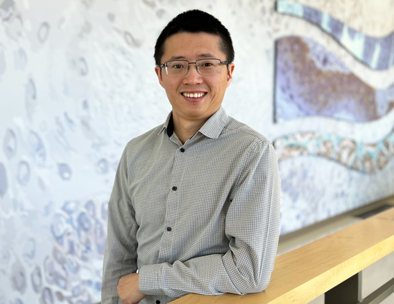
Qi Tan, PhD
Assistant Professor
- Regulatory mechanisms of tissue homeostasis in lung fibrosis. The goal of this project is to identify critical pathways through which to restore reparative or homeostatic interactions between lung resident cell types, ultimately leading to more-effective repair of the lung after injury or chronic lung diseases such as pulmonary fibrosis.
- Cell reprogramming in lung repair. Cell fate is not permanently locked in adult lung cells and can be manipulated to promote repair and regeneration. Either abnormal epithelial cells or active fibroblasts can be reprogrammed by CRISPR activation or small molecules toward reparative or homeostatic states, ultimately boosting lung repair.
- Lung organoids development. Lung organoid models developed in Dr. Tan’s lab allows recapitulation of in vivo microenvironments and maintaining cellular identity within 3D co-cultures, providing a platform for studying pulmonary fibrosis in vitro using patient-derived lung cells or iPS Cells.
- Delineating failed lung repair with next-generation sequencing. Lineage tracing of specific cell populations, and detailed analysis of their fates with RNA-seq, scRNA-seq, ATAC-seq, ChIP-seq and spatial RNA-seq, is systemically identifying the transcriptional and signaling programs that regulate lung cells during fibrosis and repair. Promising new targets are being identified and validated as key disease regulators.
Molecular Biology of Chronic Disease
Molecular Biology of Chronic Disease
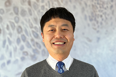
Tianshun Zhang, PhD
Assistant Professor
Dr. Zhang’s research focuses on fundamental mechanistic discoveries to prevent or treat chronic diseases. His lab’s interests are in the molecular and cellular aspects of chronic disease research, particularly in cancer signaling pathways and crosstalk between tumor and tumor microenvironment in cancer progression, metastasis, and resistance to various therapies.
Chromatin and Epigenetic Gene Regulation
Chromatin and Epigenetic Gene Regulation
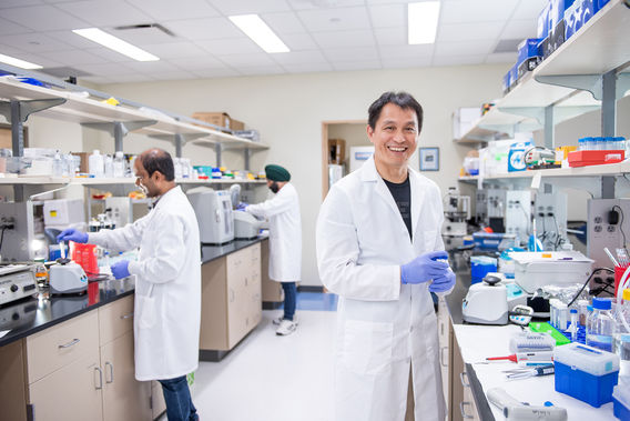
Current studies in his lab focus on elucidating the genetic and epigenetic mechanisms underlying UV-induced skin carcinogenesis and identifying actionable targets for skin cancer prevention and treatment.
Primary Research Areas
- Epigenetic mechanisms in development and cancer pathogenesis.
- Role of hairless in epidermal homeostasis and skin carcinogenesis
- New molecular targets for cancer prevention and treatment
Professional Memberships
- 2010: Society of Investigative Dermatology
- 2012: New York Academy of Sciences